


Cannabis trichomes under the electron microscope
List of contents
Cannabis flowers are consumed for medicinal and recreational purposes based on the properties of their specialised metabolites (such as cannabinoids and terpenes). The metabolites are abundantly produced within the glandular trichomes of the female flowers, which represent the essence of the prized cannabis bud. But despite the economic and medicinal importance of cannabis glandular trichomes, the relative properties and contributions of the different types of trichomes remain poorly understood.
The term trichome comes from the Greek ('thrix', 'trichos', hair or hair) and in many plant species, they form a small hairy covating. Trichome shape ranges from straight or spiral, to globular, which are those that have the greatest interest in cannabis. Trichomes perform various defensive and protective functions, and can be quite specialised; for example, the hair-like cells in carnivorous plants that trigger the trap to close on an unsuspecting insect are derived from trichomes.
Cannabis plants have a variety of trichomes, including short spiky hairs that give the leaves a rough texture and likely act as deterrents to herbivores and as an environmental buffer. Of greater historical interest, however, are the glandular trichomes, so called from the Latin 'glandis' (little acorn), due to their rounded shape.
")
Trichomes: types and growth
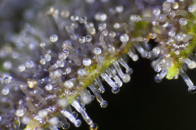
In female cannabis flowers, three types of glandular trichomes have been described based on their surface morphology: bulbous, sessile (from the Latin for 'sitting'), and stalked (or notched). Bulbous trichomes are the smallest in size and produce limited specialised metabolites. Sessile cannabis trichomes "sit" on the epidermis with a short stalk and have a globe-shaped head composed of a multicellular disc of secretory cells and a subcuticular metabolite storage cavity.
By comparison, the stalked trichomes of cannabis have a similarly shaped globe head, slightly larger, but rising several hundred microns above the epidermal surface, supported by a multicellular stalk. The relative contributions of the two main types of glandular trichomes, sessile and stemmed, to the cannabinoid and terpene profiles of cannabis flowers are still unknown. In fact, it is unclear whether pedunculate trichomes are distinct from sessile trichomes simply by the presence or absence of a stem.
 Each scale bar measures 20 µm")
Inside the cannabis bubble
To learn how these trichomes act as metabolite factories, in 2020 a Canadian research team from the University of British Columbia published a study describing how the glandular trichomes of cannabis alter their morphology and metabolite content during flower maturation. They did this by testing a CBD-rich strain of cannabis (specifically the 'Finola' hemp variety).
When we talk about molecules such as THC or CBD from the cannabis plant, we are actually talking about these glandular trichomes. Each one of those little 'balls' is a tiny factory for producing natural cannabinoids, terpenes and more, at the heart of which is a group of cells called the secretory disc: this disc sits inside the ball, just above where the rounded head meets the supporting cells of the stalk.
The Canadian research team was able to obtain remarkably clear images within these trichomes using high-resolution multiphoton microscopy, a technique that enables fluorescence emission imaging with great depth of penetration. These images showed that sessile trichomes, both on cannabis leaves and calyxes (the outer parts of the flower that protect and support it), have 8 cells in their secretory disc. Stemmed trichomes, by contrast, had significantly more cells, typically 12-16.
 and (b) are sessile trichomes of a leaf and calyx viewed from above, with each of its eight secretory cells visible. Below (c) a secretory disc of stalked (penduculated) trichomes is shown, while (d) indicates the difference in cell count between the two types.")
The study found that the additional cells in the stemmed trichomes allow for increased production of cannabinoids and other secondary metabolites. The study also found that sessile trichomes on an immature calyx (bottom left) develop into the stemmed type (right) as the plant matures. The same difference was found in a high-THC cannabis strain (specifically Purple Kush), indicating that this is not just a characteristic of CBD-rich cannabis plants.

Given their dominance in mature flowers, the psychoactive, aromatic and homeostatic abilities of cannabis that are transmitted through the glandular stalked trichomes essentially make them the key link between the plant and us.
The essence of trichomes: cannabinoids and terpenes
The techniques used in this research, which recorded the natural fluorescence of living tissue through a pulsed laser, produced an excellent visual record of the structure of cannabis trichomes. For example, in the image below, the secretory disc inside each trichome head is distinct and shaped like a loaf of bread, along with the bubble-shaped secretory cavity above the disc. This cavity swells with cannabinoids and other metabolites as the plant grows:

The bluish tint of these cells is also relevant, as the study found that the wavelengths emitted by each type of trichome are caused by their cannabinoid content. The bluish-green colour in the image above was artificially added to highlight a wavelength band from 420 to 460nm, which encompasses the maximum emission of cannabinoids. The researchers suggest that the relationship between this band of fluorescence and the longer wavelengths (495-540nm) seen in sessile leaf trichomes could be a useful diagnostic tool in the future.
But the Canadian team was not only interested in the structure of these biological formations. To understand how trichome content develops, they measured the total terpene and cannabinoid content of immature and mature plants by dissolving them in pentane; and they also used an extremely fine glass probe to extract the contents of individual trichome cavities.
Solvent extraction results showed that as the trichomes in a calyx mature, the total terpene and cannabinoid content increases. Towards the end of the plant's growth, there is also an increasing dominance of monoterpenes, the lighter type that make up essential oils and create the unique aromas of cannabis.
More specifically, the test samples showed that the increase in monoterpenes was driven by changes in stemmed trichomes. The study also looked at sessile trichomes from male plants (which are usually removed to prevent fertilization of valuable female plants) and found similarly low ratios of mono and sesquiterpenes.

The percentage of different cannabinoids in the sessile and stemmed trichomes of mature calyxes was similar, suggesting to the researchers that the sessile trichomes in this case were still converting to the stemmed type. Unsurprisingly, the non-decarboxylated form of CBD, CBDA or cannabidiolic acid, was the most prominent cannabinoid in this CBD-rich strain.
The final step the team took was to search each type of trichome for the genetic instructions that lead to the production of terpenes and cannabinoids. They found that individual glandular trichomes of all types, and in particular stem trichomes, had high levels of the genes that build both CBGA (the “parent” molecule of CBD and THC) and the specific CBDA synthase that changes CBGA to CBDA. These genes were more frequent in trichomes than in cannabis roots and shoots, as expected.
A study in microscopy that opens many doors
What does this study add to our knowledge of cannabis cultivation? Firstly, that under UV light, the stalked trichomes gave off a bright blue colour and contained a large, distinctive cake-shaped disc of cells. The smaller sessile trichomes, which are stemless, gave off a red colour, had smaller secretory discs, and produced fewer fragrant terpenes. Therefore, ultraviolet light could be used to monitor the maturity of trichomes in flowers and inform optimal harvest times.
 illuminated with ultraviolet light (f) to produce blue fluorescence from stemmed glandular trichomes to aid microcapillary sampling of resin")
Sessile and notched trichomes differ not only in that they sit on a large stalk or directly on the epidermal surface, but they also have distinct fluorescent properties, as well as differences in the number of cells in their secretory disc and terpene metabolite profiles.
Second, the unique combination of microscopic, analytical, and genetic work synthesised by the Canadian research team elucidates the growth process and molecular pathways that cannabinoids and terpenes follow, allowing greater scope to refine those processes and thus increase yields and control unwanted outcomes for medicinal or recreational cannabis users. After thousands of years of humans working with this plant, research from the University of British Columbia and their colleagues shows that we are now closer than ever to realising the full potential of this plant.
And thirdly, in reviewing scientific progress, which these days often focuses on the latest genomic breakthroughs or other big data crunches, it's easy to underestimate the value of visual aids to our understanding of the microscopic world. The detail and clarity of the images produced by this study have helped other academics, and hopefully, the general public, to understand these central generators of cannabinoids and other key molecules in cannabis.

The aesthetic value of cannabis electron microscopy
Undoubtedly, the spectacular nature of these images taken by electron microscope has seduced other researchers who have also crossed the stormy nanometric waters to show all the beauty of cannabis from a fundamental point of view. And of all the examples available in the world, Ted Kinsman's 'Cannabis: Marijuana under the Microscope' is by far the most unique.
In a market awash with books on how to grow, archives of bud porn, and explorations of cannabis culture, this work edited by this associate professor in the Department of Photographic Sciences at the Rochester Institute of Technology in New York presents weed as we'd never seen it before: through a scanning electron microscope or SEM (Scanning Electron Microscope), to get a closer look at the leaves, seeds, pollen and trichomes.

The scanning electron microscope is widely used in various fields of industry and science because it is one of the most versatile imaging and measurement tools. The photographs this technique produces are particularly prized for their great depth of field and excellent resolution, both orders of magnitude better than light microscopy. But the images provided by the SEM are black and white, and the individual images contain information in only two dimensions. However, colour is something important to us humans; and not only from an aesthetic point of view, because our brains rely on colour (and stereoscopic vision) to correctly perceive objects.
 image shows four types of trichomes on the edge of a bract leaf harvested from a large bud")
So, once Kinsman had taken the SEM images, he was able to enhance them with rich colours to make them more appealing. In fact, each image starts out black and white and must be coloured by hand. Ted paints THC-containing cannabis trichomes a bright colour to make them stand out: "I choose bold colours because I'm trying to make science visually exciting and appealing to the general population." Here is a video about Ted Kinsman's work:
Although Kinsman's book was published in 2019, back in 2014 another researcher had already planted his flag at the North Pole of cannabis images obtained through scanning electron microscopes. As you can see in another essential book: 'Cannabis Under The Microscope: A Visual Exploration of Medicinal Sativa and C. Indica' by Ford McCann, a book that also presents microscopic images of cannabis in all its glory.
As in the previous case, it is worth keeping in mind that cannabis does not actually have these bright colours like a lit-up Christmas tree. Much of the photography is in false colour because, as is the case with most images from a scanning electron microscope, computers are used to enhance and highlight certain features to make them easier to study. However, the result is still quite impressive, as seen in this example, just one of the 170 photographs in the book, and which shows that scientists, at least in the laboratory, see cannabis very differently than we growers or users do.
")
------
Images and references:
- Cannabis glandular trichomes alter morphology and metabolite content during flower maturation. Samuel J. Livingston, Teagen D. Quilichini, Judith K. Booth, Darren C. J. Wong